NADIS Sheep Disease Focus – July 2006
NADIS is a network of 40 veterinary practices and six veterinary colleges monitoring diseases of cattle, sheep and pigs in the UK. NADIS disease bulletins are written specifically for farmers, to increase awareness of prevalent conditions and promote disease prevention and control, in order to benefit animal health and welfare. Farmers are advised to discuss their individual farm circumstances with their veterinary surgeon.
NADIS data can highlight potential livestock disease and parasite incidences before they peak, providing a valuable early warning for the month ahead.
Orf (Contagious Pustular Dermatitis)
Orf is a common, highly contagious production limiting disease of sheep, goats and deer caused by a parapox virus. The disaease is characterised by proliferation of the epidermis and scab formation.
The parapox virus can survive for several months in a cool, dry environment, but is destroyed by very low temperatures, ultraviolet light and wetting. The virus may, therefore, be carried over from year to year in buildings. Sheep become infected by contact with a scab-contaminated environment or by direct contact with other infected animals. Breaks in the skin, for example associated with tooth eruption in young lambs, facilitate entry of the virus. Proliferative lesions then result from multiplication of the virus in epidermal cells, cell death and repair. The disease is self-limiting and most cases resolve within 4 – 6 weeks. Protective immunity in sheep following infection appears to be short-lived and the disease can recur, but with reduced severity. In most cases, persistently infected carrier animals are probably responsible for the annual reappearance of orf within a flock. Lambs are not protected by colostral antibodies from previously exposed dams.
Clinical signs
The first sign is redness and a slight swelling of the skin. Blisters then form, which develop into pustules which subsequently rupture to form thick scabs. After about 3 weeks, scabs are shed, leaving a layer of raw skin which heals quickly. A subsequent cycle of proliferation and repair results in the development of obvious proliferative, scab-covered lesions. Secondary bacterial infection of the lesions often occurs and scabs bleed if damaged.
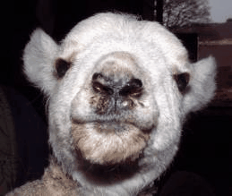
ORF LESIONS ON THE MUZZLE OF A 5 DAY-OLD LAMB
Characteristic lesions are most commonly seen on the muzzle, around the margins of the lips and around the tooth margin of the gums of 2 – 6 week-old lambs, or recently weaned lambs and hoggs. In severe cases lesions extend inside the mouth and may involve the tongue and dental pad.
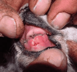
EARLY LESIONS ON THE GUMS OF A 5 DAY-OLD LAMB
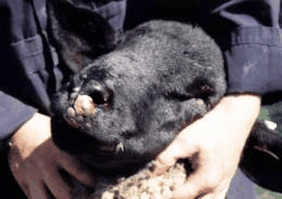
ORF LESIONS ON THE MUZZLE OF A 10 MONTH-OLD HOGG
Orf lesions are frequently seen around the base of the teats of ewes. While the disease is usually self limiting, economic losses result from failure of severely infected young lambs to suck, poor weight gains in hoggs and secondary mastitis in ewes.
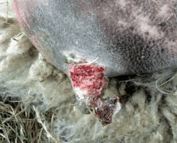
LESIONS AROUND THE BASE OF A TEAT
Orf lesions are occasionally seen at other sites such as the ears (sometimes associated with tagging); between the digits, around the coronary band and over the fetlock joints (clinically indistinguishable from ovine digital dermatitis); around the vulva of ewes; and on the penis of rams.
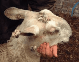
ATYPICAL ORF LESIONS ON THE EAR AND HEAD OF A BLUEFACE LEICESTER RAM
The incidence of orf within a group of sheep may exceed 50%. High levels of infection are associated with housing of January/February-born lambs, intensive stocking of recently weaned lambs, or contamination of equipment used for feeding orphan lambs. Young lambs can become infected from lesions on their dam’s teats. Scratches around the mouth caused by whins or thistles can facilitate spread of infection in hill flocks.
Orf is an important zoonosis. Painful lesions in man are usually seen on the fingers, but can occur at any site. Blisters progress to weeping sores, which persist for several weeks. The disease if often complicated by potentially serious lymphadenitis and flu-like symptoms. Human infection should be avoided by good hygiene and wearing of latex gloves when handling infected animals. The same precautions should be taken when handling the live orf vaccine.
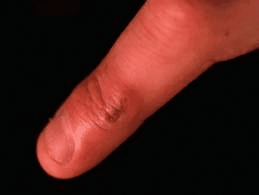
ORF LESION ON THE AUTHOR’S FINGER
Diagnosis
The diagnosis of orf is based on the clinical signs. Early lesions may resemble those of important notifiable exotic diseases, foot-and-mouth and sheep pox. The diagnosis can be confirmed by virus isolation or electron microscopy on scab material.
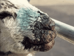
ORF INFECTION IS SOMETIMES MISDIAGNOSED AS THE CAUSE OF SEVERE PROLIFERATIVE DERMATITIS AROUD THE MUZZLE OF SHEEP GRAZED ON LUSH PASTURE UNDER VERY WET CONDITIONS. HOWEVER THE PRIMARY CAUSE IS USUALLY BACTERIAL AND MOST CASES RESPOND RAPIDLY TO ANTIBIOTIC TREATMENT
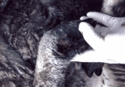
ORF VIRUS IS SOMETIMES IMPLICATED IN STRAWBERRY FOOTROT LESIONS OF THE UNDERSIDE OF THE SKIN OF THE LEGS ABOVE THE CORONARY BAND. HOWEVER THE PRIMARY CAUSE IS USUALLY DERMATOPHILUS INFECTION ASSOCIATED WITH PERSISTENT WETTING
Treatment
There is no proven treatment for orf. There is little evidence to show any beneficial effect of capsules which are marketed for orf treatment. Furthermore administering these capsules presents a high risk for human infection. Oxytetracycline aerosol sprays can be applied to control secondary infection, which may reduce the severity of the lesions. Severely infected animals may also benefit from parenteral antibiotic injections. Provision of palatable, soft, supplementary feed is important in cases where severe mouth lesions are present.
Prevention
Orf can be managed by vaccination. In most flocks, the preferred methods are to vaccinate housed early lambs as they are turned out of lambing pens and older lambs at weaning. Alternatively, ewes can be vaccinated at least 8 weeks before lambing, with the aim of reducing the level of infection at lambing from teat lesions. The vaccine is live, so it is important that sufficient time elapses, for scabs to heal, between vaccination of the ewes and lambing. Vaccination can also be used to protect in contact sheep in flocks where the disease has appeared. Vaccination immunity only lasts for about 6 – 8 months and vaccination of ewes affords no colostral protection to lambs. The vaccine strain of the virus can produce disease, so its use should not be considered in flocks with no history of orf.
Application is by scratching the skin twice with a scarifying needle dipped in vaccine.
Young lambs can be vaccinated on the skin between the top of the foreleg and the chest wall, where they are unable to lick. Older lambs are usually vaccinated inside the thigh and ewes on the skin under the tail. The prongs of the applicator should be wiped regularly to remove any build up of grease. The vaccine is live, so should not come into contact with disinfectants and animals should not be scratched so vigourously as to make them bleed. The scratch sites of a few animals should be inspected after about one week for evidence of rows of small pustules, which indicate that vaccination has been effective. Vaccines should be kept out of direct sunlight, refrigerated at 2 – 8o C and used before the expiry date.
The severity of outbreaks of orf can be reduced by avoiding overcrowding, or contact with whins or thistles, although these are seldom practical options.
Copyright © NADIS 2006 www.nadis.org.uk
| FURTHER INFORMATION | SPONSORS’ LINK |
| • Supporting British Livestock click here |  |
| FURTHER INFORMATION | SPONSORS’ LINK |
| • To find out more about EBLEX, click here |
| FURTHER INFORMATION | SPONSORS’ LINK |
| • To find out more about the HCC click here |
| FURTHER INFORMATION | SPONSORS’ LINK |
| • To find out more about QMS click here |


