

Why ultrasound scanner is one vet’s favourite bit of kit
 © Wyvern Farm Vets
 If I had to choose one piece of kit I wouldn’t want to be without as a vet, would it be the traditional yet still indispensable stethoscope or thermometer?
Or cutting-edge technology such as the blood-sampling machine that diagnosis liver disease in less than 10 minutes?
I think it would have to be the ultrasound scanner, because it provides immediate answers without being invasive.
This makes it one of the most valuable diagnostic tools available to farm vets.
See also: Benefits of lung scanning for respiratory disease in calves
Ultrasound scanners send high-frequency sound waves, produced using tiny vibrating crystals from the handheld probe, that travel through tissue and record the bounce back, to create a real-time image of what’s happening inside the animal.
These sound waves can move through muscle, tissue and fluid but not through air.
Reproductive performance
For most farmers, ultrasound scanning is synonymous with pregnancy diagnosis.
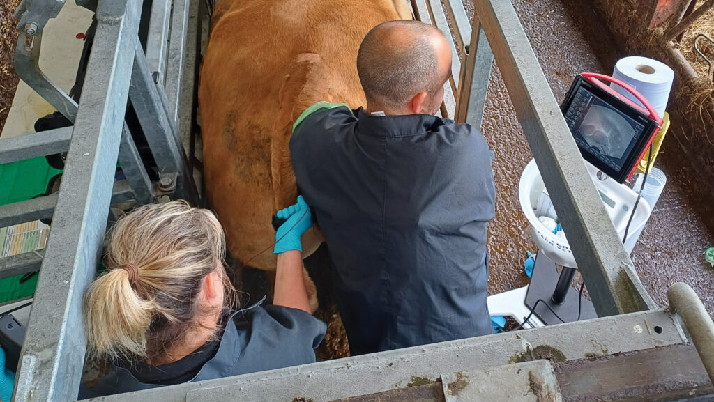
During routine fertility visits, vets can use the scanner to produce images of the internal structures within and around the uterus by passing the probe rectally and sending sound waves below through the rectal and uterine walls.
The scanner allows us to examine the uterus and ovaries, confirming whether a cow is pregnant, while also assessing whether the embryo is alive and developing normally.

Lewis Wosnitzka © Wyvern Farm Vets
We can estimate the stage of pregnancy, identify twins and, at the appropriate stage, even determine the sex of the calf.
Just as importantly, ultrasound helps us manage fertility before pregnancy occurs.
By examining the ovaries, we can identify follicles, corpus luteum and ovarian cysts, giving us valuable information about where a cow is in her breeding cycle.
Improving reproductive performance has significant benefits for both profitability and animal health.
Better fertility means shorter calving intervals, fewer days open and improved lifetime productivity, all of which contribute to a more efficient dairy or beef enterprise.
Early detection of calf pneumonia
Ultrasound has also transformed the way we diagnose pneumonia in young calves.
Traditionally, calves were only treated once obvious clinical signs such as coughing, high temperatures or poor appetite had developed. Unfortunately, by this stage, lung damage has often already occurred.
Lung ultrasound allows vets to detect pneumonia much earlier. By scanning the lungs through the ribs, we can visualise the integrity of the pleural surface of the lungs and identify lesions that indicate early pneumonia.
Early diagnosis means treatment can begin sooner, often reducing disease severity, improving recovery and supporting more responsible antibiotics use with fewer retreatments recorded.
Similarly, skilled vets can use the scanner to visualise the heart and check for congenital abnormalities, abnormal function or hardware disease.
The long-term benefits are considerable. Research has shown that heifers experiencing calfhood bovine respiratory disease had an average daily gain reduced by 0.067kg/day and produced 121.2 kg less milk during their first lactation (Buczinski et al, 2021), highlighting the lasting impact of early disease.
Long-term benefits
The scanner continues to prove its worth throughout the cow’s life.
It can be used to assess the liver when investigating metabolic disease, examine the uterus after calving for signs of infection and investigate heart conditions. It can even assess the teat and udder alongside milk culture when investigating mastitis cases.
While ultrasound technology has advanced enormously, its greatest strength remains the speed with which it provides information.
Instead of relying on guesswork, vets and farmers can make evidence-based decisions immediately, allowing treatment to be targeted where it will have the greatest benefit.